

Biological imaging ranges from the visualization of ions, molecules, cells and tissues to whole small animal models, and is one of the most used and demanded technologies in life science related research, teaching and industrial applications. The BF-BioImaging platform supports, maintains, and develops a powerful portfolio of various state-of-the-art equipment and expertise in a wide range of light and electron microscopy services, including light microscopy modalities for both fixed samples and live cell and tissue imaging, ranging from super-resolution to light sheet and mesoscopic imaging, and from label-free imaging to high-content microscopy. Electron microscopy offerings cover standard and cryo-genic specimen preparation for transmission and scanning electron microscopy, three-dimensional imaging at wide length scales and various hybrid techniques with light microscopy.
The platform comprises over 50 full-time RI personnel in 12 imaging units organized into 5 nodes in Helsinki, Turku, Oulu, Tampere, and Kuopio. The platform serves a large user base of >1400 users, has contributed to several patents, start-ups, and company collaborations, and supports other BF technology platforms by providing various microscopy-based readouts and analysis tools.
BF-BioImaging platform services include:
- Open access to all facilities
- Personal user support and general support activities: e.g., consultation on experimental set up; sample preparation; choice of instruments and imaging methods; image analysis; user training & guidance
- Maintenance and development of imaging systems, methods and analytics
- Teaching activities: lecture and practical courses for MSc & graduate students & postdoctoral fellows
- Imaging data management
- Support in image analysis and development of image analysis tools
- International activities
Nodes
UEF: University of Eastern Finland; UH: University of Helsinki; UO: University of Oulu; UTU: University of Turku; TAU: Tampere University; ÅAU: Åbo Akademi University
Contact details
Platform Chair
Eija Jokitalo
eija.jokitalo@helsinki.fi
Platform Vice Chair
Pasi Kankaanpää
pkanka@utu.fi
Services
Helsinki BioImaging (HBI)
Biomedicum Imaging Unit – Light Microscopy (BIU-LM) provides access to advanced widefield, confocal and super-resolution light microscopy as well as a wide selection of image post-processing and analysis tools. Specialized technologies include several super-resolution modalities, light-sheet and fluorescence lifetime microscopy.
BI Light Microscopy Unit (LMU-BI) provides access, training and consultation on basic light microscopy techniques such as widefield, high-content and confocal microscopy, as well as advanced techniques including super-resolution, fluorescence lifetime and light-sheet/light field microscopy. Cell culture facilities, as well as a wide range of image analysis tools and services are also provided.
FIMM High Content Imaging and Analysis (FIMM-HCA) provides access and service in (confocal) high-content (HC) imaging, including automation with high throughput plate handler and cell incubator. Specialized technologies include computer assisted laser microdissection for phenotypic isolation of single cells for further omics analytics. Expertise in HC image processing and advanced analysis is offered.
BI Electron Microscopy Unit (EMBI) provides wide range of sample preparation techniques and access to transmission and scanning electron microscopy and image analysis tools. The unit specializes in advanced volume EM workflows comprising serial block face SEM, focused ion beam SEM, array tomography, correlative light-electron microscopy and methods for image segmentation and analysis.
Turku BioImaging (TBI)
Turku BioImaging Operations Team (TBI-OT) provides open access to advanced image analysis and data management services, including OMERO servers and deep learning solutions. TBI-OT also coordinates imaging services and activities in Turku, and the national coordination between BF- BioImaging, Euro-BioImaging Finland and Euro-BioImaging ERIC.
The Advanced Imaging Core (AIC-Turku) provides access, training, and consultation across a wide range of light microscopy modalities. The facility is equipped with widefield, spinning-disk, and point-scanning confocal microscopes, as well as light-sheet imaging and super-resolution platforms including Airyscan, STED, and single-molecule localization microscopy (SMLM). Due to its location within the Turku Bioscience Centre, users can readily combine light microscopy with complementary core facilities including cytometry, zebrafish, and spatial and single-cell omics.
The Laboratory of Electron Microscopy (TBI-EM) provides standard sample preparation and access to transmission electron microscopy. TBI-EM offers advanced techniques such as immuno electron microscopy, electron tomography and correlative light-electron microscopy.
Oulu BioImaging (OBI)
Light Microscopy Core Facility (OBI-LM) provides access to point-scanning and spinning-disk confocal microscopy, including total internal reflection fluorescence (TIRF), fluorescence recovery after photobleaching (FRAP), and fluorescence lifetime imaging (FLIM). For larger fixed and living samples, the facility offers light-sheet microscopy, optical projection tomography (OPT), multiphoton microscopy with lifetime and spectral imaging, and fluorescence stereomicroscopy. Optical clearing services are available, and a dedicated intravital imaging laboratory supports imaging of anesthetized mice using multiphoton microscopy with a resonant scanner and full surgical and physiological monitoring equipment.
Electron Microscopy Core Facility (OBI-EM) provides sample preparation and access for transmission and scanning electron microscopes, specializes in mouse phenotyping, immuno electron microscopy, and offers advanced 3D Serial imaging on SEM (Array tomography) and correlative light-electron microscopy techniques.
Tampere BioImaging (TAUBI)
Tampere Imaging Facility (TIF) specializes in imaging of cell cultures, tissues, small model organisms (e.g., zebrafish), (bio)materials, light-responsive materials, and 3D cell cultures and organoids. The facility hosts a wide range of imaging systems, including several widefield fluorescence microscopes, five confocal microscopes, a total internal reflection fluorescence (TIRF) microscope, a TCSPC-based fluorescence lifetime imaging microscope (FLIM), and a super-resolution structured illumination microscopy (SR-SIM) system.
Eastern Finland BioImaging (EFBI)
Cell and Tissue Imaging Unit (CTIU) provides access, services, and consultation in imaging, experimental design, and image analysis. The unit is equipped with high-content imaging platforms for live-cell imaging, confocal microscopes for high- and super-resolution imaging and molecular dynamics with incubators for long-term time-lapse experiments, a TCSPC-based FLIM microscope for quantitative analysis of live and fixed samples, and a widefield fluorescence microscope with a slide scanning stage and computational clearing. A histology laboratory supporting sample processing is also available.
Electron microscopy core facility (SIB Labs) provides sample preparation and access for transmission and scanning electron microscopies, elemental analysis and specializes in multimodal imaging combining EM and spectroscopy.
Recent user publications
Ershov et al. TrackMate 7: integrating state-of-the-art segmentation algorithms into tracking pipelines (2022) Nature Methods 19:829- ; PMID: 35654950; DOI: 10.1038/s41592-022-01507-1
Devarajan et al. Targeting collagen XVIII improves the efficiency of ErbB inhibitors in breast cancer models (2023) Journal of Clinical Investigation 133:e159181; PMID: 37498672; DOI: 10.1172/JCI159181
Kyykallio et al. A quick pipeline for the isolation of 3D cell culture-derived extracellular vesicles (2022) Journal of Extracellular Vesicles 11:e12273; PMID: 36257915; DOI: 10.1002/jev2.12273
Danielsson et al. Nuclear lamina strain states revealed by intermolecular force biosensor (2023) Nature Communications 14: 3867- ; PMID: 37391402; DOI: 10.1038/s41467-023-39563-6
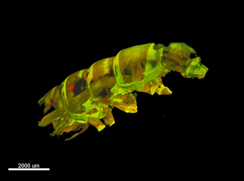
Kirstin Vonderstein, Light sheet microscopy, Caterpillar.
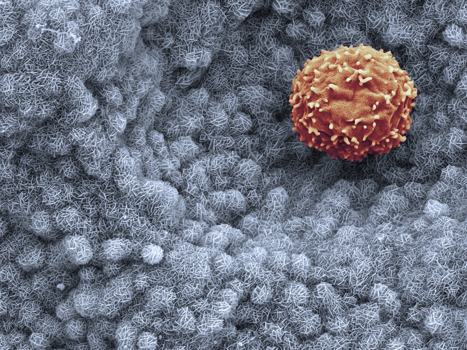
Ilkka Miinalainen: Waiting for differentiation.
